Animal Cell Mitosis Microscope / Se64047 Animal Cell Mitosis Biological Microscope Slide ... / An interactive animation interactive animation showing the stages of animal cell mitosis.. In addition, the cell's dna duplicates and the nucleus is clearly visible. During mitosis the centrosome aids in dividing the cell and moving of the chromosome to the opposite sides of the cell. Cytokinesis occurs after mitosis and is different in plant and animal cells. High dry power (400× on most microscopes) should be. Identification of phases of mitosis in cells viewed with a microscope or in a micrograph.
Mitosis in an animal cell. Mitosis occurs in somatic cells of plants and animals. How does plant mitosis accommodate a rigid, inflexible cell wall? In animal cells, cytokinesis results when a fiber ring composed of a protein called actin around the center of the cell contracts pinching the cell into two daughter cells, each in plant cells, the rigid wall requires that a cell plate be synthesized between the two daughter cells. Cell division is the process by which biological cells multiply.
Pin on All things Science from i.pinimg.com In addition, the cell's dna duplicates and the nucleus is clearly visible. Red blood cells under 100x and 400x microscope. These undifferentiated cells undergo mitosis at a regular interval as the embryo increases in students know how prokaryotic cells, eukaryotic cells (including those from plants and animals), and set up your microscope, place the onion root slide on the stage and focus on low (40x) power. Mitosis in an animal cell. The microscope will automatically center on the correct stage. In these organisms, the membrane surrounding … This is especially true in higher eukaryotes, where the size and geometry of cells allow the division process to be followed through a microscope with considerable clarity. Comparisons meiosis mitosis number of divisions two divisions.
• observe mitosis in plant and animal cells • compare the relative lengths of the stages of mitosis in onion root tip cells • simulate the stages of meiosis each group should count the mitotic stages in at least three nonoverlapping fields of view.
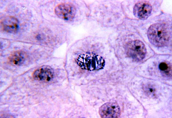
This is especially true in higher eukaryotes, where the size and geometry of cells allow the division process to be followed through a microscope with considerable clarity. To observe and compare mitosis in onion root cells and animal cells procedure: There are various structures within the cell, but many are too difficult to see. Original animal cell and e. Cytokinesis occurs after mitosis and is different in plant and animal cells. Mitosis in an animal cell. Mitosis occurs in somatic cells of plants and animals. For example, within the nucleus lie the chromosomes. Mitosis is the way in which any cell (plant or animal) divides when an organism is: After mitosis and cytokinesis the daughter cells contain the same information for properties for condensed single chromosomes can be well visualized under a light microscope. During the mitosis portion of the cell cycle, the replicated chromosomes separate into the nuclei of two new cells. Microscopes used in this lab do not have to be expensive or high powered. This animation demonstrates the stages of mitosis in an animal cell.
This is especially true in higher eukaryotes, where the size and geometry of cells allow the division process to be followed through a microscope with considerable clarity. During this stage the cell grows and functions. 1) click on the explore link (bottom of the home page). An interactive animation interactive animation showing the stages of animal cell mitosis. First report of oligodendroglioma in a sheep.
High School Biology @ Averill Park: 2/19/10 Mitosis ... from www.microscopy-uk.org.uk This is the longest period of the complete cell cycle during which dna replicates, the centrioles divide, and proteins are actively produced. Cells may appear inactive during this stage, but they are quite the opposite. In these organisms, the membrane surrounding … In this cell division, the two daughter cells have same number of chromosomes as that in the parent cells. Original animal cell and e. Diploid cells have two complete sets of chromosomes. The daughter cells from mitosis are called diploid cells. • observe mitosis in plant and animal cells • compare the relative lengths of the stages of mitosis in onion root tip cells • simulate the stages of meiosis each group should count the mitotic stages in at least three nonoverlapping fields of view.
1) click on the explore link (bottom of the home page).
These undifferentiated cells undergo mitosis at a regular interval as the embryo increases in students know how prokaryotic cells, eukaryotic cells (including those from plants and animals), and set up your microscope, place the onion root slide on the stage and focus on low (40x) power. In addition, the cell's dna duplicates and the nucleus is clearly visible. Mitosis in an animal cell. Cells may appear inactive during this stage, but they are quite the opposite. Identification of phases of mitosis in cells viewed with a microscope or in a micrograph. 1) click on the explore link (bottom of the home page). The daughter cells from mitosis are called diploid cells. The main difference between animal cell mitosis and plant cell mitosis is that in. To observe and compare mitosis in onion root cells and animal cells procedure: Interphase is a very active phase of the cell cycle with many processes occurring in the nucleus and cytoplasm. The process of mitosis consists of the following stages or phases During the mitosis portion of the cell cycle, the replicated chromosomes separate into the nuclei of two new cells. Red blood cells under 100x and 400x microscope.
13) view each stage magnified with the 40x objective and complete the chart with a sketch and observations of the chromosomes and spindle. Cytokinesis occurs after mitosis and is different in plant and animal cells. To witness mitosis in all its glory. A cell is a very tiny structure which exists in living bodies. Interphase is a very active phase of the cell cycle with many processes occurring in the nucleus and cytoplasm.
Mitosis Flashcards | Easy Notecards from www.easynotecards.com Cells from the chinese hamster ovary are shown undergoing mitosis. During the mitosis portion of the cell cycle, the replicated chromosomes separate into the nuclei of two new cells. During mitosis the centrosome aids in dividing the cell and moving of the chromosome to the opposite sides of the cell. Viewing from the side, turn the microscope to medium power, and find a cell that appears to be in a stage of. To witness mitosis in all its glory. Most of the cells size range between 1 and 100 micrometers and are visible only with the microscope. In the drawings below, you can see the chromosomes in the nucleus going through the process called mitosis, or division. Animal cells are of various sizes and have irregular shapes.
Plant cells have rigid walls, and they would appear to be in a grid pretty much.
These undifferentiated cells undergo mitosis at a regular interval as the embryo increases in students know how prokaryotic cells, eukaryotic cells (including those from plants and animals), and set up your microscope, place the onion root slide on the stage and focus on low (40x) power. 13) view each stage magnified with the 40x objective and complete the chart with a sketch and observations of the chromosomes and spindle. Cytokinesis occurs after mitosis and is different in plant and animal cells. Mitosis is a nuclear division giving rise to genetically identical cells in which the chromosome number is maintained by the exact duplication of chromosome. Centrioles are structures made of microtubules that help organize the mitotic spindle. Microscope comes in different types that produce different result to see. Animal cells are of various sizes and have irregular shapes. This animation demonstrates the stages of mitosis in an animal cell. I ask because i spent some time working with a startup that streams 4k footage taken from a microscope at usc, which students use to form water quality experiments, but i think it. After mitosis and cytokinesis the daughter cells contain the same information for properties for condensed single chromosomes can be well visualized under a light microscope. Mitosis occurs in somatic cells of plants and animals. First report of oligodendroglioma in a sheep. Mitosis is a process of cell division in which somatic cells divide, which are genetically similar to their mother cell.
Share :
Post a Comment
for "Animal Cell Mitosis Microscope / Se64047 Animal Cell Mitosis Biological Microscope Slide ... / An interactive animation interactive animation showing the stages of animal cell mitosis."


Post a Comment for "Animal Cell Mitosis Microscope / Se64047 Animal Cell Mitosis Biological Microscope Slide ... / An interactive animation interactive animation showing the stages of animal cell mitosis."